Ultrastructure and cellular organization of longsnout seahorse Hippocampus reidi liver
DOI:
https://doi.org/10.47193/mafis.3912026010108Keywords:
Hepatocyte, electron microscopy, histological morphologyAbstract
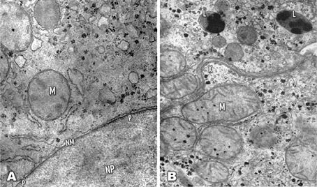
Hippocampus reidi is an endangered tropical species, as well as being one of the most widely traded marine fish globally. The cellular organization of the normal liver in the longsnout seahorse (H. reidi) was studied using electron microscopy to provide an overview of the liver’s fine structure. Two hundred and fifty seahorses were collected and prepared for transmission electron microscopy. Sections were cut using an LKB ultramicrotome and then washed with PBS, stained with uranyl citrate for 2-3 min, and then with lead acetate for 20 s. The grids were then observed using an electron microscope Zeiss EM 109. The cytoplasm of the hepatocyte contains a nucleus surrounded by a double membrane, diffusely scattered mitochondria and a network of the endoplasmic reticulum of rough and smooth types. Most of the rough endoplasmic reticulum appear to be in parallel to the nuclear envelope or the cell membrane and was situated relatively in the center part of the cytoplasm. The cellular organization of the longsnout seahorse liver is quite similar to that of other mammalian species.
Downloads
References
Aschoff L. 1924. Das reticulo-endotheliale system. In: Kraus F, Meyer E, Minkowski O, Müller F, Sahli,H, Schittenhelm A, Czerny A, Heubner O, Langstein L, editors. Ergebnisse der inneren medizin und kinderheilkunde. Ergebnisse der inneren medizin und kinderheilkunde. Vol 26. Berlin: Springer. p. 1-118.
[CITES] Convention on International Trade in Endangered Species of Wild Fauna and Flora. 2009. Appendices II listing proposal for Hippocampus. https://www.cites.org/eng/resources/species.html.
Foster S, Wiswedel S, Vincent A. 2016. Opportunities and challenges for analysis of wildlife trade using CITES data-seahorses as a case study. Aquat Conserv Mar Freshwat Ecosyst. 26 (1): 154-172.
Freret-Meurer NV, Andreata V. 2008. Field studies of a Brazilian seahorse population, Hippocampus reidi Ginsburg, 1933. Braz Arch Biol Technol. 51 (4): 743-751.
Furth R, Cohn ZA, Hirsch JG, Humphrey JH, Spector WG, Langevoort HL. 1972. The mononuclear phagocyte system: a new classification of macrophages, monocytes, and their precursor cells. Bull WHO. 46 (6): 845-852.
Gingerich WH. 1982. Hepatic toxicology of fishes. In: Weber LJ, editor. Aquatic toxicology. New York: Raven Press. p. 55-105.
Gupta S. 2000. Hepatic polyploidy and liver growth control. Semin Cancer Biol. 10 (3): 161-171.
Hinton DE, Pool CR. 1976. Ultrastructure of the liver in channel catfish Ictalurus punctatus (Rafinesque). J Fish Biol. 8: 209-219.
Hora MSC, Joyeux J-C, 2009. Closing the reproductive cycle: growth of the seahorse Hippocampus reidi (Teleostei, Syngnathidae) from birth to adulthood under experimental conditions. Aquaculture. 292: 37-41. DOI: https://doi.org/10.1016/j.aquaculture.2009.03.023
Ito T. 1973. Recent advances in the study on the fine structure of the hepatic sinusoidal wall: a review. Gunma Rep Med Sci. 6: 119-163.
Koning S, Hoeksema BW. 2021. Diversity of seahorse species (Hippocampus spp.) in the international aquarium trade. Diversity. 13 (5): 187. DOI: https://doi.org/10.3390/d13050187
Naito M, Hasegawa G, Takahashi K. 1997. Development, differentiation, and maturation of Kupffer cells. Microsc Res Tech. 39: 350-364. DOI: https://doi.org/10.1002/(SICI)1097-0029(19971115)39:4<350::AID-JEMT5>3.0.CO;2-L
Oliveira T, Pollom R. 2017. Hippocampus reidi. The IUCN Red List of Threatened Species 2017: e.T10082A17025021.
Yokota S, Fahimi HD. 1981. Immunocytochemical localization of albumin in the secretory apparatus of rat liver parenchymal cells. Proc Nat Acad Sci USA. 78 (8): 4970-4974. DOI: https://doi.org/10.1073/pnas.78.8.4970
Smedsrod B, de Bleser PJ, Braet F, Lovisetti P, Vanderkerken K, Wisse E, Geerts A. 1994. Cell biology of liver endothelial and Kupffer cells. Gut. 35: 1509-1516. DOI: https://doi.org/10.1136/gut.35.11.1509
Smedsrod B, Pertoft H, Eggertsen G, Sundstrom C. 1985. Functional and morphological characterization of cultures of Kupffer cells and liver endothelial cells prepared by means of density separation in Percol, and selective substrate adherence. Cell Tissue Res. 241: 639-649.
Widmann JJ, Cotran RS, Fahmi HD. 1972. Mononuclear phagocytes (Kuffer cells) and endothelial cells: identification of two functional cell types in rat liver sinusoids by endogenous peroxidase activity. J Cell Biol. 52: 159-170. DOI: https://doi.org/10.1083/jcb.52.1.159
Published
Issue
Section
License
Copyright (c) 2025 Luis A. Romano, Luana B. Giesta, Virginia F. Pedrosa, Michael H. Schwarz, Luís A. Sampaio, Ricardo V. Rodrigues

This work is licensed under a Creative Commons Attribution-NonCommercial-ShareAlike 4.0 International License.
Authors of articles published in Marine and Fishery Sciences retain copyright on their articles, except for any third-party images and other materials added by Marine and Fishery Sciences, which are subject to copyright of their respective owners. Authors are therefore free to disseminate and re-publish their articles, subject to any requirements of third-party copyright owners and subject to the original publication being fully cited. Visitors may also download and forward articles subject to the citation requirements. The ability to copy, download, forward or otherwise distribute any materials is always subject to any copyright notices displayed. Copyright notices must be displayed prominently and may not be obliterated, deleted or hidden, totally or partially.
This journal offers authors an Open Access policy. Users are allowed to read, download, copy, distribute, print, search, or link to the full texts of the articles, or use them for any other legal purpose within the Creative Commons 4.0 license (BY-NC-SA), without asking prior permission from the publisher or the author. This is in accordance with the BOAI definition of Open Access.